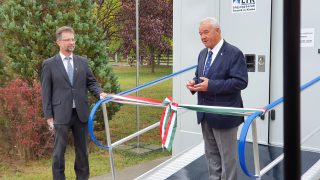
With strict adherence to the epidemiological measures, a small handover ceremony of the standing equine MRI device took place on 7th of October 2020, which arrived at the Department and Clinic of Equine Medicine of the University of Veterinary Medicine Budapest.
Dr Péter Sótonyi, Rector of the University, welcomed the guests, members of the Marek József Foundation’s board of trustees, Dr Gábor Náray-Szabó president, Dr István Bajkai, Dr Sándor Fazekas, Dr Balázs Aladics, as well as Dr Gyula Budai, Ministerial Commissioner, the representatives of Hungarian equestrian sports and horse breeders, vice-rectors and colleagues of the university.
Professor Péter Sótonyi rector began his opening speech in remembrance of the past by recalling the victim of the thirteen Martyrs of Arad executed on October 6, 1849, after the fall of the Hungarian Civic Revolution and War of Independence. Sótonyi highlighted that the Department and Clinic of Equine Medicine have recently undergone several investments and renovations, which will continue in the future.
After the renovation of the clinical department and the set up of the isolation boxes, a new generation MRI device arrived in September, which represents a next step towards providing the highest level of veterinary care for horses and to those competing in equestrian sports.
“This is a huge opportunity for us,” stressed Dr Péter Sótonyi rector – since with the arrival of the new MRI, not only a device became  available at our university, but we also have the appropriate intellectual sources to operate it. The development of the Clinic does not stop here, in the near future we plan to purchase a CT, an ultrasound and tooth monitoring device, as well as to build a complex system connecting and operating all of our diagnostic devices.
The operation of the new device was described by Dr Annamária Nagy, the specialist in charge of the MRI, who came home from England at the call of the university, where he worked with a similar device for more than a decade. With the standing equine MRI, it is possible to examine the foot, pastern, fetlock, metacarpal/metatarsal, carpal and tarsal regions. It provides detailed information on the soft tissue and bone patterns of the limbs, as well as allows the diagnosis of lesions which is not visible by ultrasound without the unnecessary risk of general anaesthesia.
After the speeches, the MRI device was handed over with a ribbon-cutting ceremony.
Click here for the photo gallery.
Click here to learn more about the MRI device and the examination process.
