Lameness is one of the most frightening terms for the equestrian society. Not only because the pain of the animal is clearly visible, but also the origin and cause of this type of disease is often difficult or impossible to determine. If the diagnosis is not reached by conventional diagnostic tools such as X-ray or ultrasound, you may consider other advanced imaging modalities such as magnetic resonance imaging (MRI), which has recently arrived at the Department and Clinic of Equine Medicine of the University of Veterinary Medicine Budapest.
About MRI
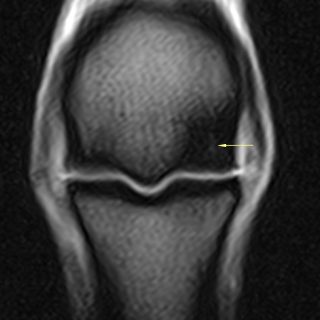
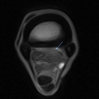
MRI has been used in human medicine for decades. This highly reliable diagnostic imaging modality can take detailed pictures of a specific area of the body, helping to make an accurate diagnosis and plan the most effective therapy. MRI has been available for horses since the 1990s, and a device that can examine horses in a standing position without the unnecessary risk of general anaesthesia has been used since 2002. This new generation device can detect lesions that remain invisible to X-rays or ultrasounds. Another advantage is that bone and soft tissue structures can be examined simultaneously. With the help of the standing equine MRI, it is possible to examine the foot, pastern, fetlock, metacarpal/metatarsal, carpal and tarsal regions.
Highly recommended
It is advisable to use MRI if: a negative or inconclusive result is obtained on X-ray or ultrasound if the problematic area cannot be reached with ultrasound (for example lesions in the hoof capsule) if the chosen treatment fails or the animal responds poorly to it. It can also be used to verify the treatment and check the recovery before the animal returns to work or competition, and it can also help to detect fractures in racehorses at an early stage.
About the MRI scan
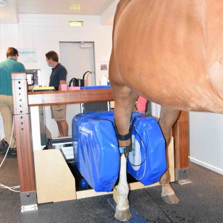
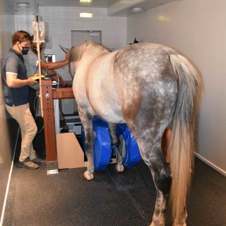
The first step is to localize the lameness with diagnostic anaesthesia, and then we can start to prepare the horse for the MRI scan. The shoes must be removed prior to MRI examination because the presence of metal in the magnetic field can compromise image quality. MRI is performed in a standing sedated horse. A catheter is inserted into the horse’s jugular vein, through which a sufficient amount of sedation is given frequently in small doses to the animal. After that, the horse enters the room where the MRI machine is located, and the limb is placed in a U-shaped magnet. The procedure ideally takes about two hours. After the examination, the horse is put in a stable until the effect of intoxication is completely gone – which usually means a few hours – so in the vast majority of cases, the horse can go home on the day of the examination. During an MRI scan, hundreds of images are taken of a given area of the body, which are evaluated by a specialist veterinarian. The result is sent to the owner of the horse and the referring veterinarian within 24 hours.
To treat lameness effectively, it is recommended to act as soon as possible, because early and accurate diagnosis and treatment can prevent worsening of the injury, further injuries due to compensation for pain, and can significantly reduce the time of recovery.
The modern standing equine MRI, which arrived at the Department and Clinic of Equine Medicine of the University of Veterinary Medicine Budapest, has created a unique opportunity in Central and Eastern Europe for horse owners and those interested in equestrian sports. With the new diagnostic tool and the team of specialists, it can be stated without exaggeration that health care for horses can now be provided at a world-class level.


